Tracking vascular remodelling in post-ischemic stroke
Ischemic stroke, Diabetic mouse model, 2-photon imaging
Ischemic stroke is a leading cause of disability, with profound impacts on neural and vascular networks in affected brain regions such as the cortex. Understanding how cellular and vascular remodeling occurs post-stroke is critical for developing targeted rehabilitation strategies.
Using 2-photon imaging , we were able to track chronic changes in blood flow and vasculature structure, before and after ischemic stroke in a diabetic mouse model. The goal of this project is to further develop robust machine-learning algorithms and imaging processing toolkits to analyze the large datasets generated from these imaging sessions. These tools will enable us to systematically quantify changes in blood cell density, aggregate movement, and vascular restructuring. In addition, we will investigate whether interventions like pharmacological treatments can mitigate brain damage and promote recovery in the post-stroke brain. By creating this toolkit, this research aim to establish a potential biomarker for disease diagnosis and the evaluation of rehabilitation strategies.
Using 2-photon imaging , we were able to track chronic changes in blood flow and vasculature structure, before and after ischemic stroke in a diabetic mouse model. The goal of this project is to further develop robust machine-learning algorithms and imaging processing toolkits to analyze the large datasets generated from these imaging sessions. These tools will enable us to systematically quantify changes in blood cell density, aggregate movement, and vascular restructuring. In addition, we will investigate whether interventions like pharmacological treatments can mitigate brain damage and promote recovery in the post-stroke brain. By creating this toolkit, this research aim to establish a potential biomarker for disease diagnosis and the evaluation of rehabilitation strategies.
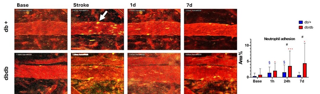

2024 Sato Y, Li Y, Kato Y, Kanoke A, Sun JY, Nishijima Y, Wang RK, Stryker M, Endo H, Liu J. Type 2 diabetes remodels collateral circulation and promotes leukocyte adhesion following ischemic stroke. Journal of Cerebral Blood Flow and Metabolism (in print) doi:10.1101/2024.10.23.619748
pmc.ncbi.nlm.nih.gov/articles/PMC11526934/
pmc.ncbi.nlm.nih.gov/articles/PMC11526934/